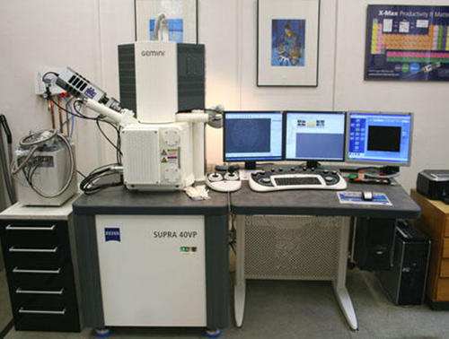
Scanning electron microscope (SEM) specializing in paleontology
The scanning electron microscope provides microphotographs with a magnification of up to five thousand times. The images generated are images of the object surfaces and have a high depth of field.
The scanning electron microscope provides microphotographs with a magnification of up to five thousand times. The images generated are images of the object surfaces and have a high depth of field. Objects from micropaleontology such as foraminifera, ostracods and gastropods are routinely analyzed using this instrument.
High-resolution imaging methods provide an essential research basis in all areas of work in the field of paleontology. A scanning electron microscope is therefore an essential element of basic equipment. The increasing comparative inclusion of recent organisms and their ultrastructural conditions makes a device with low beam acceleration and high signal and contrast yield necessary (field emission) in order to avoid radiation through thin organic layers on the one hand and distorting conductivity coatings on the one hand. The electrical charges, which can hardly be avoided, especially in cavernous objects, can be counteracted with the help of the VP mode (Variable Pressure).
Model:
ZEISS SUPRATM 40 VP Ultra with a thermal field emission cathode (with variably adjustable pressure for detailed topographic imaging of non-conductive samples) and Oxford Instruments EDX system (X-ray analysis) with INCA analysis software.
Accesories:
Sputtering device (Gold) - Biorad Polar Division
Contact
Jan Evers
Room D.032
+49 (0)30 - 838 70 246
email: j.evers@fu-berlin.de
